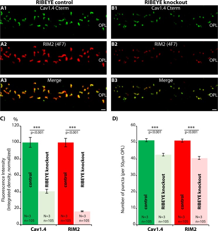
Figure 9.
Confocal analyses of photoreceptor synapses from RIBEYE knockout (B) and littermate control retinas (A) immunolabelled with mouse monoclonal antibodies against RIM2 (4F7) and rabbit polyclonal antibodies against Cav1.4 (Cav1.4 Cterm). (A,B) Representative confocal images, (C,D) quantitative analyses of the strength of the respective immunosignals (integrated density in C) and number of immunoreactive puncta (in D) plotted as mean ± S.E.M. Abbreviations: OPL, outer plexiform layer. Scale bars: 1 μm (A–D).

