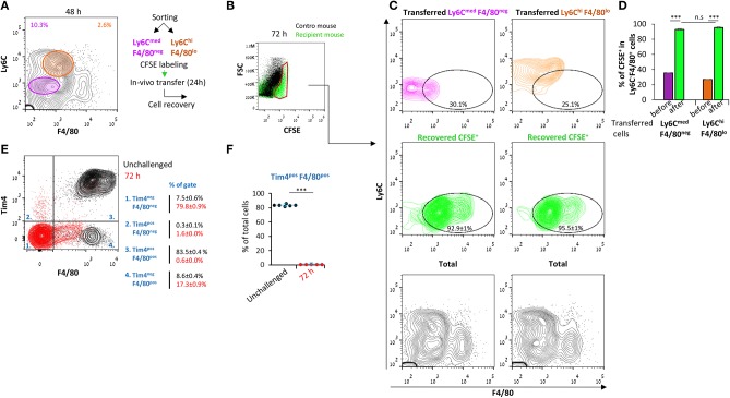
Figure 3.
Ly6CmedF4/80neg and Ly6ChiF4/80lo cells both convert to Ly6Cneg F4/80+ macrophages. Peritoneal exudates were recovered from WT mice 48 h PPI. (A) Monocytic cells were sorted into Ly6CmedF4/80neg and Ly6ChiF4/80lo populations. (B–D) Sorted cells were labeled with CFSE and transferred to recipient mice with ongoing peritonitis at 48 h. At 72 h, peritoneal cells were recovered, immunostained for Ly6C and F4/80, and CFSE+ cells (B) were analyzed by flow cytometry (C, D). Results are stacked contour plot from six mice (C) and means ± SEM (n = 6). P < 0.001 (Student's t-test). (E,F) Peritoneal exudates were recovered from unchallenged mice or at 72 h PPI, immunostained for F4/80 and Tim4 and analyzed by flow cytometry. Results are stacked contour plots from six mice (E) and percentage means ± SEM of F4/80+ Tim4+ cells (F).***P < 0.001(Student's t-test).

