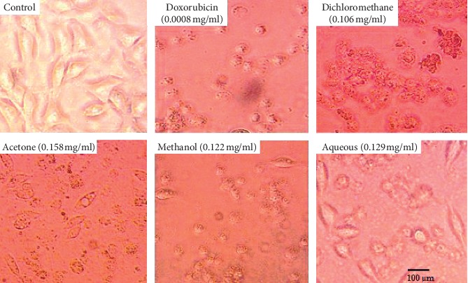
Figure 1.

Morphological alterations on RD cells at IC50 of SAHA extracts and doxorubicin. Photomicrographs taken at 400x magnification using a Japson MD130 digital camera. The pictures were analyzed using a future win joe software for windows. Untreated RD cells (control) appeared in normal shape with 95–100% confluence. RD cells treated with SAHA extracts and doxorubicin showed loss of cell adherence, shrinking of cells, and reduced cell density along with cell debris.
