
Figure 1.
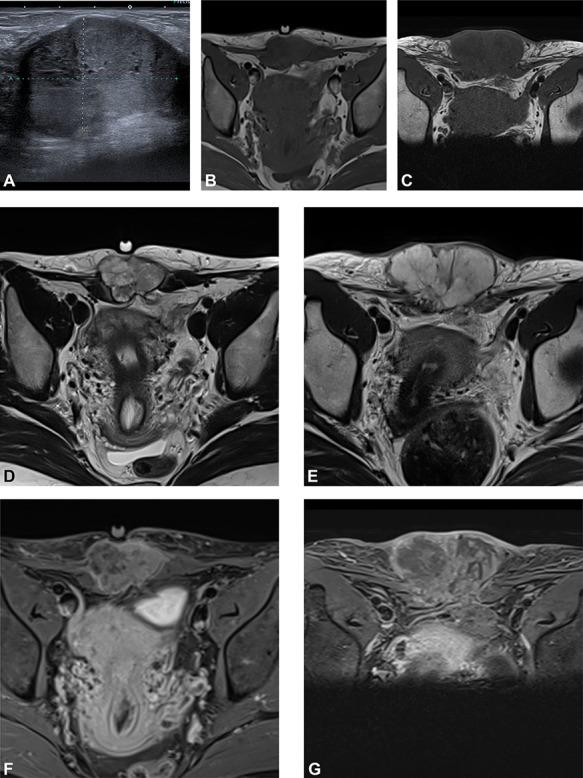
(A) Preoperative ultrasound: ultrasound exam at diagnosis showing a well-defined mass with acoustic enhancement and diffuse ground-glass echoes. (B) Magnetic resonance imaging (MRI) findings: before neoadjuvant radiotherapy (B, D, F) and at the end of the treatment (C, E, G). MRI exams show a well-defined solid mass which presents iso-signal on T1WI (B, C), hyper signal on T2WI (D, E) and heterogeneous enhancement after gadolinium injection (F, G). This mass partially involves both rectus abdominis muscles. A progression, mainly due to necrosis, with an increased size after radiotherapy can be observed.