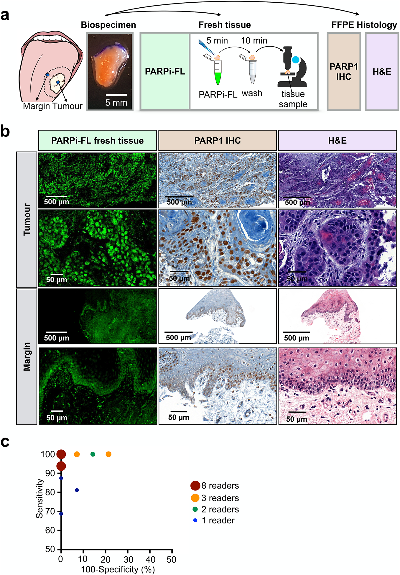
Fig. 5 |. Rapid PARPi-FL staining on fresh biospecimens.

a, Workflow of rapid PARPi-FL staining of fresh human biospecimens. Biospecimens were split and separately processed for fresh tissue staining as well as histopathology. b, Examples of PARPi-FL staining of a tumour and margin sample and corresponding histopathology of the same sample. We aimed to scan the entire fresh tissue in a high-resolution tile scan. Lower and higher magnification images showcase PARPi-FL staining and corresponding PARP1 expression in the specimens. A total of 22 tissues (n=12 tumours and n=10 benign tissues adjacent to tumour from 13 individual patients) were stained and analysed. The full data set is provided in supplementary Materials PDF files 1 and 2. c, In a blinded study, readers (n=27) scored 30 cases (n=12 tumours, n=10 margins, n=8 duplicates (4 tumours, 4 margins)) as tumour or margins (see fig. S9 for study design). Pairs of sensitivity and specificity for each reader are represented in the graph.
