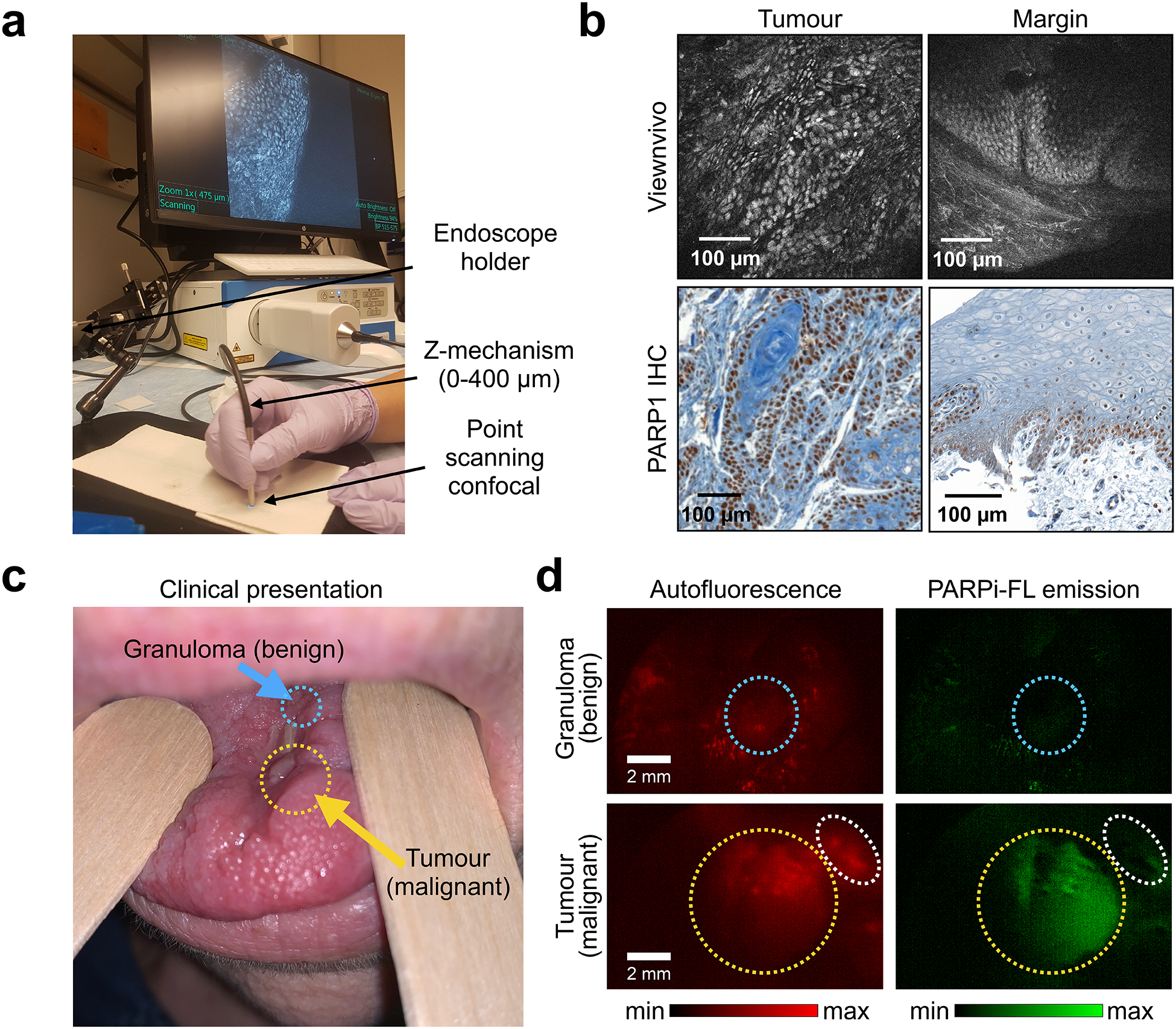
Fig. 6 |. Feasibility of microscopic and macroscopic in-human imaging of PARPi-FL.

a, Imaging setup for the ViewnVivo point scanning confocal endomicroscope suitable for in vivo imaging. b, Fluorescence images from a patient sample stained with 100 nM PARPi-FL and corresponding PARP1 IHC. c, PARPi-FL first-in-human imaging (NCT03085147), showing a patient who presented with a malignant recurrent squamous cell carcinoma (yellow arrow) and an adjacent benign granuloma (blue arrow). d, Using a Quest Spectrum imaging device with a laparoscopic camera and PARPi-FL optimized laser/filter system, the tumour area of the patient was imaged after gargling a 500 nM PARPi-FL solution. Granuloma (blue circles) and tumour (yellow circles) areas showed distinct patterns of PARPi-FL accumulation, and specific PARPi-FL uptake was only detectable in the tumour. The white circles highlight a non-malignant region which showed autofluorescence, but no PARPi-FL uptake.
