Figure 3.
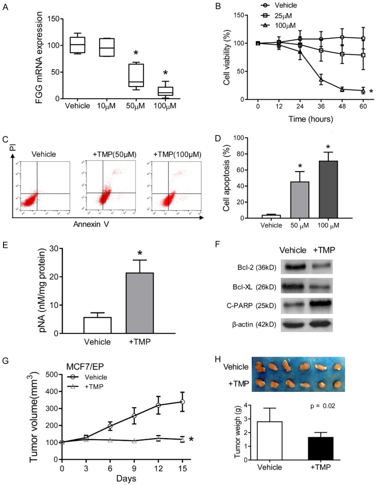
TMP reduces FGG expression and reverses anthracycline resistance of breast cancer cells. (A) MCF7/EP cells were treated with a gradient concentration of TMP for 48 h. The mRNA levels of FGG were measured with qRT-PCR assays. β-actin was used as control. (B) Cell viability of MCF7/EP cells were detected with CCK-8 assays. MCF7/EP cells were treated with combined EPI (3 μM) and indicated concentration of TMP for 60 h. (C) MCF7/EP cell apoptosis were measured in different treatment: EPI (3 μM) + vehicle, EPI (3 μM) + TMP (50 or 100 μM). Cell apoptosis was analyzed with flow cytometry by Propidium iodine and Annexin V staining. (D) Statistics of MCF7/EP cell apoptosis in combined treatment or EPI only groups as described in (C). (E) Activity of caspase-3 was measured by pNA concentrations with Caspase-3 Activity Assay Kit. (F) Apoptosis related proteins (cleaved PARP, Bcl-2 and Bcl-xL) in TMP treated MCF7/EP cells were analyzed with Western blot assays. β-actin was used as loading control. (G) Xenografts growth of MCF7/EP cells with the treatment of EPI with/without TMP. Tumor volume was recorded after the treatment with the mean tumor volumes in the indicated day. (H) Xenografts were harvested and imagined after 15 days’ treatment. The tumor weights were compared between groups. Bar = 1 cm. Data represent the means ± SD. *P < 0.05.

