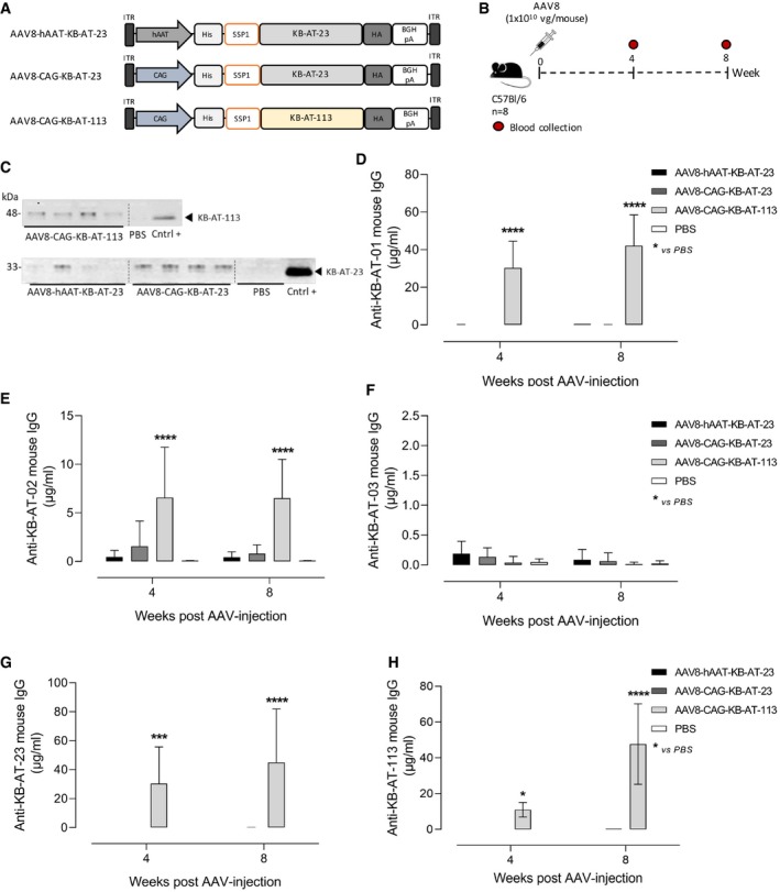
Figure 7. Assessment of sdAbs potential immunogenicity in wt mice.

-
ASchematic representation of the AAV8 vectors used to express KB‐AT‐23 and KB‐AT‐113 variants. ITR: inverted terminal repeats for AAV packaging.
-
BScheme representing the study design. Vectors were injected in C57Bl/6 mice (n = 8) at a dose of 1E+10 vg/mouse. Red symbols represent timing of blood collection.
-
CRepresentative Western blot analyses on plasma samples (n = 4) collected from animals 4 weeks post‐AAV injection. Cntrl +: positive controls represented by the loading of the purified KB‐AT‐23 or KB‐AT‐113 proteins.
-
D–HMeasurement of anti‐sdAb mouse IgG in plasma samples collected at days 28 and 57 post‐AAV injection. The ELISA plate was coated with the purified KB‐AT‐01 (D), KB‐AT‐02 (E), KB‐AT‐03 (F), KB‐AT‐23 (G), or KB‐AT‐113 (H) sdAbs. Data are reported as mean ± SD (n = 8/group). Data were analyzed in a 2‐way ANOVA with Dunnett's correction of multiple comparisons. *P < 0.05, ***P < 0.01 ****P < 0.001.
Source data are available online for this figure.
