Figure 5.

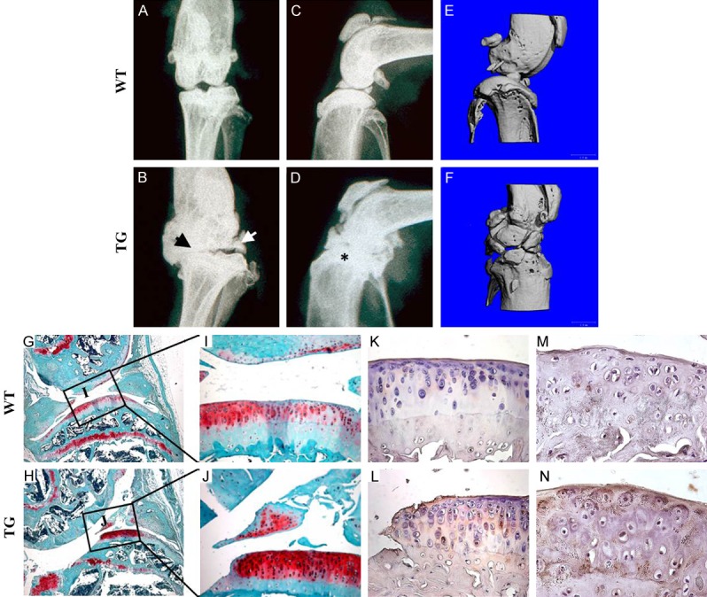
Spontaneous osteoarthritis development in eleven-month-old transgenic mice: (A-D) Radiographs of the knee joints of eleven-month old TG and WT mice. Note the characteristic osteoarthritis features in the TG mice: joint space narrowing (B, black arrow), development of osteophytes (B, white arrow), and sub-chondral sclerosis (D, asterisk). (E, F) The micro-CT analysis of the knee joints of eleven-month-old TG and WT mice revealed rougher bone surfaces and numerous osteophytes protruding into the joint cavity of the TG mice, but not WT mice. (G-J) SO/FG staining of the knee joints of the eleven-month-old TG and WT mice. Note the absence of Safranin-O staining at the articular surfaces in TG mice. (K, L) Type X collagen immunostaining revealed the ectopic Col10a1 expression in the articular cartilages of TG mice, but not of WT mice. (M, N) Immunostaining showed elevated secretion of matrix metalloprotease-13 (Mmp13) in the articular cartilages of 11 months old TG mice.
