Figure 1.
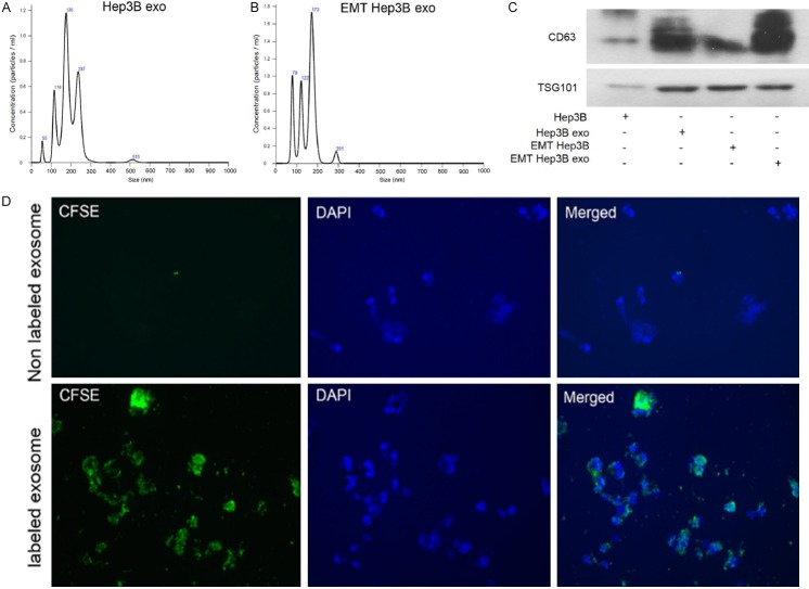
Exosomes secreted by Hep3B and TGF-β1-induced Hep3B were taken up by Hep3B cells. A and B. Purified particles were analyzed by nanoparticle tracking analysis (NTA). Sizes of the particles were between 50 and 200 nm. C. Expression of exosome markers (CD63 and TSG101) was detected by western blot. The protein expression of exosome markers was upregulated in Hep3B-exo and EMT-Hep3B exo compared with their cells. D. CFSE-labeled Hep3B-exosomes showed green fluorescence in the cytoplasm of Hep3B cells. EMT Hep3B exo: exosomes secreted from Hep3B induced by TGF-β1.

