Figure 1.

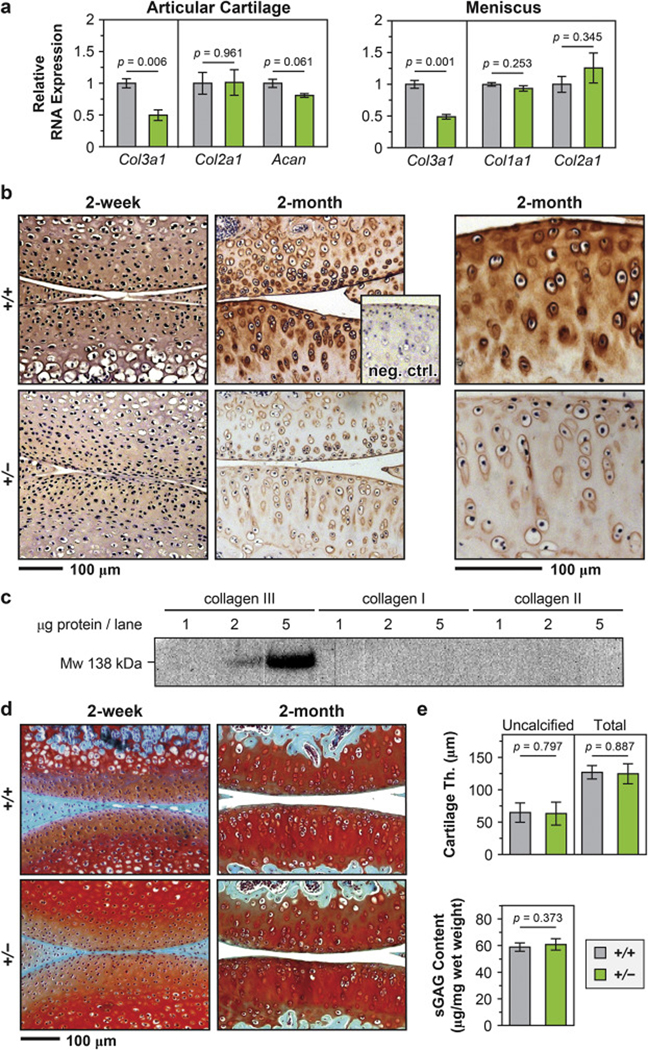
Distribution of collagen III in articular cartilage and the impact of collagen III on cartilage gross-level morphology. a) Quantitative PCR (qPCR) showed that in Col3a1+/– (+/–) mouse, the expression of Col3a1 gene was significantly reduced by ≈ 50% in both articular cartilage and meniscus (n = 4 for wild-type (+/+), n = 3 for +/−, mean ± SEM, p < 0.01), while other major matrix genes were not significantly affected (Col2a1, Acan in articular cartilage and Col1a1, Col2a1 in meniscus). b) Immunohistochemistry (IHC) of collagen III in murine articular cartilage did not show clear distribution pattern of collagen III at 2-week age, but detected intense localization in the PCM at 2-month age, and reduced staining in Col3a1+/– cartilage. Shown together is the negative internal control of WT cartilage stained without primary antibody. c) Western blot on recombinant human collagen I, III and collagen II extracted from human articular cartilage validated the specificity of the antibody for collagen III, AB7778. d) Safranin-O/Fast Green histology illustrated no appreciable differences in the staining of sGAGs or joint morphology between +/+ and +/− mouse knee articular cartilage at both 2-week and 2-month ages. e) No significant differences were found in the thicknesses of uncalcified or total cartilage and the amount of sGAGs between 2-month-old +/+ and +/− cartilage (mean ± 95% CI, n ≥ 6 for each genotype).
