Fig. 1.
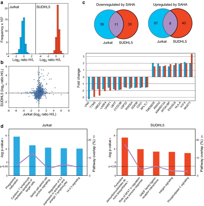
a Raw distribution of SILAC quantifications given as log2 (heavy (control)/light (SAHA) ratios. b Scatterplot of the normalized SILAC data showing protein expression changes (log2 scale) in Jurkat (x-axis) and SUDHL5 (y-axis) after SAHA treatment. c Venn diagrams illustrating significant differentially expressed proteins in Jurkat and SUDHL5 after SAHA treatment (upper panel). The lower panel shows the 19 common proteins that were significant differentially (> 1.5-fold) expressed in the two cell lines (linear scale). d DEPs from Jurkat (left) and SUDHL5 (right) were presented to IPA analysis. The most confidently affected pathways are shown with -log p values on the Y-axis, indicated with a line is p = 0.05. The percentage of overlap of the pathway is also denoted

