Fig. 3.
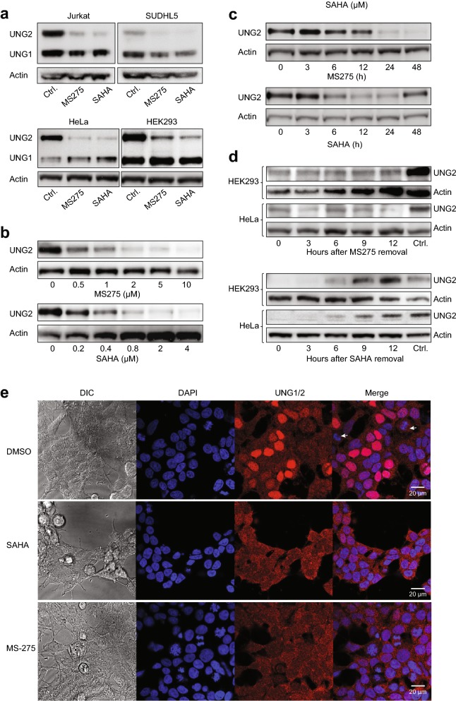
HDAC inhibition mediates selective depletion of the nuclear UNG2 isoform in various cell lines. a Western analysis of TCEs from Jurkat, SUDHL5, HeLa and HEK293 cells using a polyclonal antibody (PU059) recognising the common catalytic domain of nuclear UNG2 and mitochondrial UNG1. The membranes were subsequently probed with anti-actin antibodies as loading controls. Robust depletion by both 5 µM MS-275 and 2 µM SAHA was specific for the UNG2 isoform. b HEK293 cells were treated with various concentrations of SAHA or MS-275 for 24 h, and TCEs were subject to western analysis as in a. c HEK293 cells were treated with 2 µM SAHA or 5 µM SAHA, harvested at different time points and subject to western analysis as in a. d Induction of UNG2 expression after HDACi removal. After 18 h culture of HEK293 and HeLa cells in media containing 5 µM MS-275 (top panels) or 2 µM SAHA (bottom panels) with optimal depletion, cells were cultured in media without HDACi and UNG2 expression monitored by western analysis at the given time points. e DAPI nuclear staining (blue) and immunocytochemical staining of UNG1/2 (red) in HEK293 cells treated with 2 µM SAHA, 5 µM MS-275 or DMSO vehicle for 24 h prior to immunostaining with polyclonal PU59 antibody. Note the selective depletion of nuclear UNG2 whereas mitochondrial UNG1 remains unaffected after HDACi treatment. The differential UNG2 staining in the DMSO controls reflects the strict cell-cycle dependent expression of the nuclear UNG2 isoform, which peaks in late G1/S and is lowest in G2 and M-phase (white arrows indicate mitotic cells). DIC; Differential interference contrast images of the same sections

