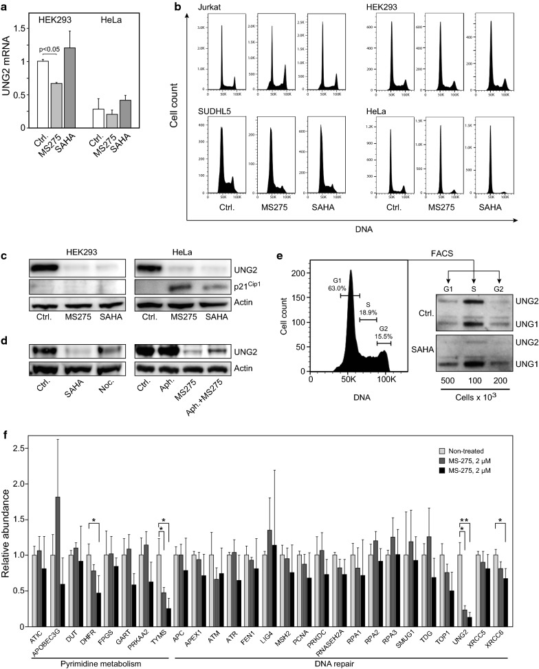
Fig. 4.
a Quantitative RT-PCR of UNG2 mRNA isolated from HeLa and HEK293 cells treated with 2 µM SAHA, 5 µM MS-275 or DMSO for 24 h. b Flow cytometry histograms of HEK293-, HeLa-, Jurkat- and SUDHL5 cells treated with 2 µM SAHA, 5 µM MS-275 or DMSO for 24 h. c Expression of p21Cip1 in HEK293- (left) and HeLa cells (right) treated as in c. d Treatment of HEK293 with cells 2 µM SAHA for 24 h mediated a stronger depletion of UNG2 than the G2/M-blocking agent nocodazole (10 µM, 24 h) (left panels). MS-275 mediated robust inhibition of UNG2 in cells arrested in G1/S by co-treatment with aphidicolin (10 µM, 24 h) (right panels). e HEK293 cells were treated with 2 µM SAHA for 12 h and subjected to fluorescence-activated cell sorting into G1-, S- and G2 fractions. Percentages of cells in each cell cycle phase are indicated in the flow cytometry histogram (left panel) and western blots showing UNG2 expression in SAHA- and DMSO -treated cells (right panels). f Expression of 27 selected proteins involved in pyrimidine metabolism as well as DNA repair were quantified by PRM. Each bar represents the mean of at least three biological replicates with SDs as indicated (* p < 0.05). Proteotypic peptides employed in the PRM analyses are given in Additional file 6: Table S2

