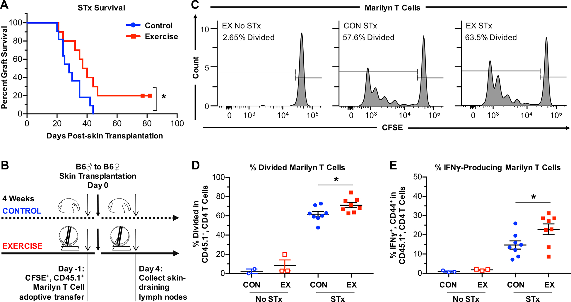
Figure 2. Exercise results in prolonged allograft survival.

(A) Rejection kinetics of minormismatched skin allografts in control (n=11) or exercised (n=10) female B6 mice. Survival curve comparisons were performed using Mantel-Cox test. (B) Experimental design to examine priming in the dLNs using H-Y–specific, CD45.1+ congenic, CD4+ Marilyn T cells. Marilyn T cells were labeled with CFSE and transferred (2×10⁵ or 6×10⁵ cells/mouse) into control or exercised female B6 mice 1 day prior to transplantation with male B6 skin grafts. Mice were sacrificed 4 days after transplantation, and cells were isolated from the graft dLNs (axillary, brachial, and inguinal) for analysis of CFSE dilution. (C) Representative plots of CFSE dilution and divided Marilyn T cells. (D) Quantitation of divided Marilyn T cells in Marilyn-gated T cells on day 4 post-transplantation. (E) Percentage of IFNγ+ cells among Marilyn T cells after restimulation with anti-CD3 and anti-CD28 for 24 hours. (B-E) CON = control, EX = exercise, No STx = no skin transplantation, STx = skin transplantation from male B6 mouse. (D-E) Each point represents a single mouse. Results are displayed as mean ± SEM. Comparisons were performed using a two-tailed unpaired t test. *p<0.05. Results were combined from 2 independent experiments.
