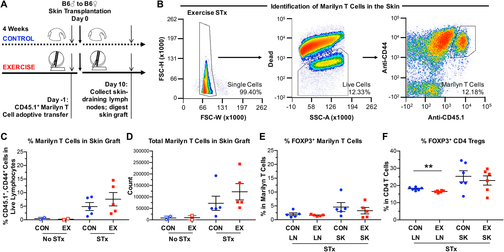
Figure 3. Exercise does not reduce T cell infiltration into the graft.

(A) Experimental design to examine T cell recruitment into the allograft using H-Y–specific, CD45.1+ congenic, CD4+ Marilyn T cells. Marilyn T cells were transferred (2×10⁵ cells/mouse) into control or exercised female B6 mice 1 day prior to transplantation with male B6 skin grafts. Mice were sacrificed 10 days after transplantation, and cells were isolated from both the dLNs and the skin graft. For mice that did not receive a skin graft, shaved flank skin was harvested. (B) Gating strategy for the identification of Marilyn T cells in the skin. Single cells were pre-gated on lymphocytes and Marilyn T cells identified as CD44hi, CD45.1+ cells. (C) Percentages and (D) total numbers of CD44hi, CD45.1+ cells (Marilyn T cells) in the skin allograft at 10 days post-transplantation, shown normalized per gram of skin graft. (E) Percentages of Foxp3+ cells among CD44hi, CD45.1+ cells (Marilyn T cells) in both the dLNs and skin allograft at 10 days post-transplantation. (F) Percentages of Foxp3+ cells among bulk CD4+ T cells in both the dLNs and skin allograft at 10 days post-transplantation (p=0.0059). (C-F) LN = draining lymph nodes, SK = skin allograft. Each point represents a single mouse. Results are displayed as mean ± SEM. Comparisons were performed using a two-tailed unpaired t test. **p<0.01. Results were combined from 2 independent experiments.
