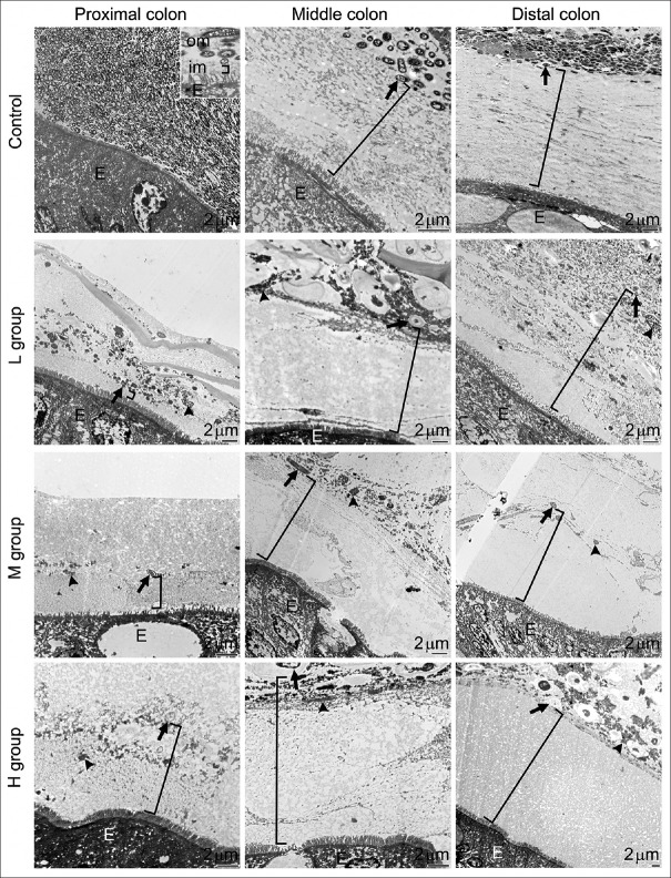
Figure 3.
Effects of antibiotics on the intestinal mucus layer. These representative transmission electron microscopy images show the proximal, middle, and distal colon taken from control (upper panel) mice and mice treated with 0.5 g (second panel, L group), 1.0 g (third panel, M group), or 1.5 g (fourth panel, H group) of SB. The inner mucus layer (im, bracket symbols in all images) is clearly identifiable in the images from control colons: A relatively homogeneous and electron-lucent area spanning between the epithelium (E) and the bacteria-rich outer mucus layer (om). The thickness of the inner mucus layer significantly increased from the proximal to the distal colon in control mice. Antibiotics effectively decreased bacterial numbers and only a few remained morphologically intact (arrows) in the outer mucus layer. The increased concentrations of electron-dense materials (arrowheads) in the outer mucus layer are bacterial debris

