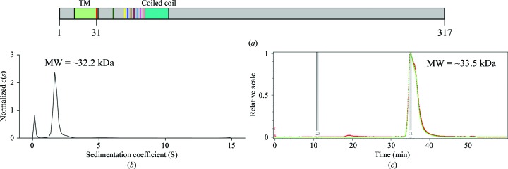
Figure 1.
Characterization of Amuc_1100*. (a) Predicted diagram of Amuc_1100. The transmembrane region (TM) and coiled-coil domain are colored light green and cyan, respectively. Residues 31–317 are the protein construct used in this experiment. Different truncated versions are indicated in various colors. (b) Analytical ultracentrifugation (AUC) analysis of Amuc_1100*. The horizontal axis is the sedimentation coefficient and the vertical axis is the normalized c(s). The molecular weight of Amuc_1100* is approximately 32.2 kDa. (c) Static light-scattering (SLS) results. The horizontal axis represents time and the vertical axis represents the relative scale. The molecular weight of Amuc_1100* is approximately 33.5 kDa.

