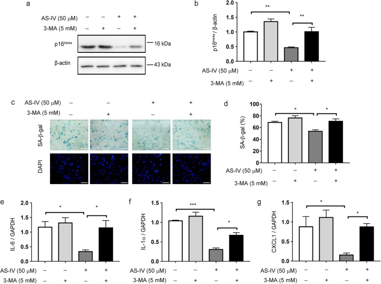
Fig. 6.
Inhibition of autophagy reverses the suppressive effect of AS-IV on astrocyte senescence. Astrocytes (40 days) were treated with 3-MA (3 mM) for 1 h before AS-IV (50 μM) treatment for 10 days. a, b Representative immunoblots (a) and quantitative analysis of p16Ink4a (b) in astrocytes. Quantified data are normalized to the control group (the control group value is equal to 1). c, d Representative images of SA-β-gal activity (c) and the percentage of SA-β-gal+ cells (d). DAPI staining nucleus (blue). Scale bar 100 μm. e–g qPCR of IL-6 (e), IL-1α (f), and CXCL1 (g) mRNA levels. The data shown are the mean ± SEM from three to five independent experiments. *p < 0.05, **p < 0.01, ***p < 0.001. AS-IV, Astragaloside IV

