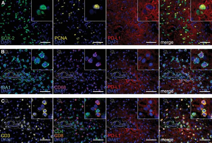
FIGURE 4.
Multiplex immunofluorescence of the tumor microenvironment in post-treatment glioblastoma with inflammatory response to checkpoint inhibition. A, Immunolabeling with the nuclear stem cell marker SOX-2 (green), proliferation marker PCNA (yellow), and PD-L1 (red; SP142). PD-L1/SOX-2/PCNA+ cells are depicted in the merged image. B, Representative images of perivascular immune cells co-expression of IBA1 (aqua), CD68 (purple), and PD-L1 (red). C, Representative images of T-cells (CD3, yellow) showing co-expression of both CD4 helper T-cells (green) and CD8 cytotoxic T cells (orange) with PD-L1 (red). Nuclei are counterstained with 4',6-diamidino-2-phenylindole (DAPI; blue). Scale bar = 50 μm.

