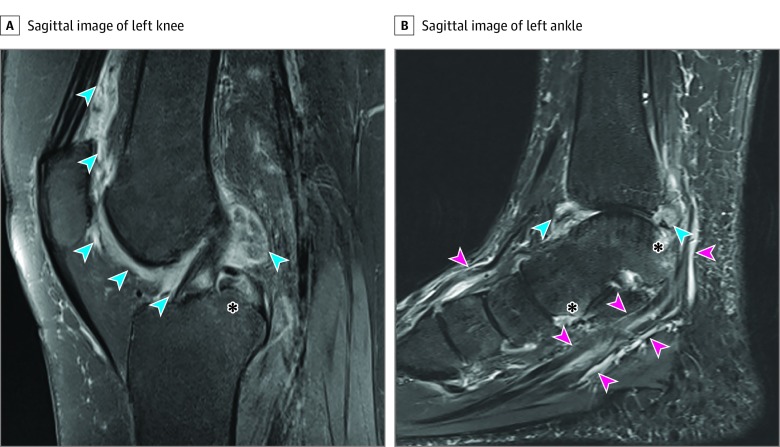
Figure 1. Magnetic Resonance Image of the Knee and Ankle of an Individual With Cervical Cancer and Immune Checkpoint Inhibitor–Induced Inflammatory Arthritis (Patient 7).
A, Sagittal fat-suppressed, proton density fast spin-echo image of the left knee depicts extensive, irregular synovial thickening (blue arrows) at the anterior and posterior aspects of the knee and bone marrow edema (asterisk). B, Sagittal image of the left ankle from a short tau inversion recovery sequence demonstrating synovial thickening at the tibiotalar joint (blue arrows), tenosynovitis (pink arrows), and periarticular bone marrow edema (asterisk).

