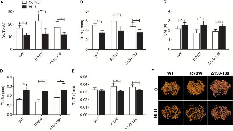
FIGURE 1.
Femoral trabecular bone loss caused by 4 weeks of HLU in WT and Cx43 transgenic mice. μCT analysis reveals decreases in BV/TV (A) and Tb.N (B) and increases in SMI (C) and Tb.Sp (D); Tb.Th (E) is decreased in mutant but not in WT mice. Comparisons via two-way with Bonferroni test (A–E). Data represent the means ± SD. n = 6–10/group. *P < 0.05, **P < 0.01, ***P < 0.001. (F) μCT images of femoral trabecular microstructure. HLU, hindlimb unloading; WT, wild-type; μCT, micro-computed tomography; R, R76W; Δ, Δ130–136; BV/TV, bone volume fraction; Tb.N, trabecular number; SMI, structure model index; Tb.Sp, trabecular separation; Tb.Th, trabecular thickness.

