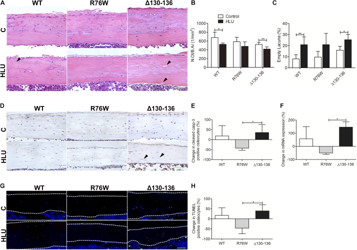
FIGURE 3.
Changes in osteocyte survival in the tibial cortical bone of Cx43 transgenic mice following HLU. (A) Representative hematoxylin-eosin-stained tissue sections of tibia cortical bone; solid arrowheads indicate empty lacunae. Osteocyte number (N.Ot/B.Ar) is decreased (B) and number of empty lacunae is increased (C) in both WT and Δ130–136 mice following HLU. n = 5–7/group. Decreased cleaved caspase-3 (D,E) and TUNEL signal (G,H) are observed in R76W mice but not in the other groups. n = 3/group. (F) Real-time PCR shows significantly decreased caspase 3 expression in R76W compared with Δ130–136 mice in the cortical bone. White dashed lines indicate bone margins. n = 3/group. Scale bar = 60 μm. Comparisons via two-way with Bonferroni test (B,C) or one-way ANOVA with Turkey test (E,F,H). Data represent the means ± SD. *P < 0.05, **P < 0.01. HLU, hindlimb unloading; WT, wild-type; R, R76W; Δ, Δ130–136; B.Ar, bone area; BS, bone surface; C, control; N.Ot, osteocyte number; B.Ar, bone area.

