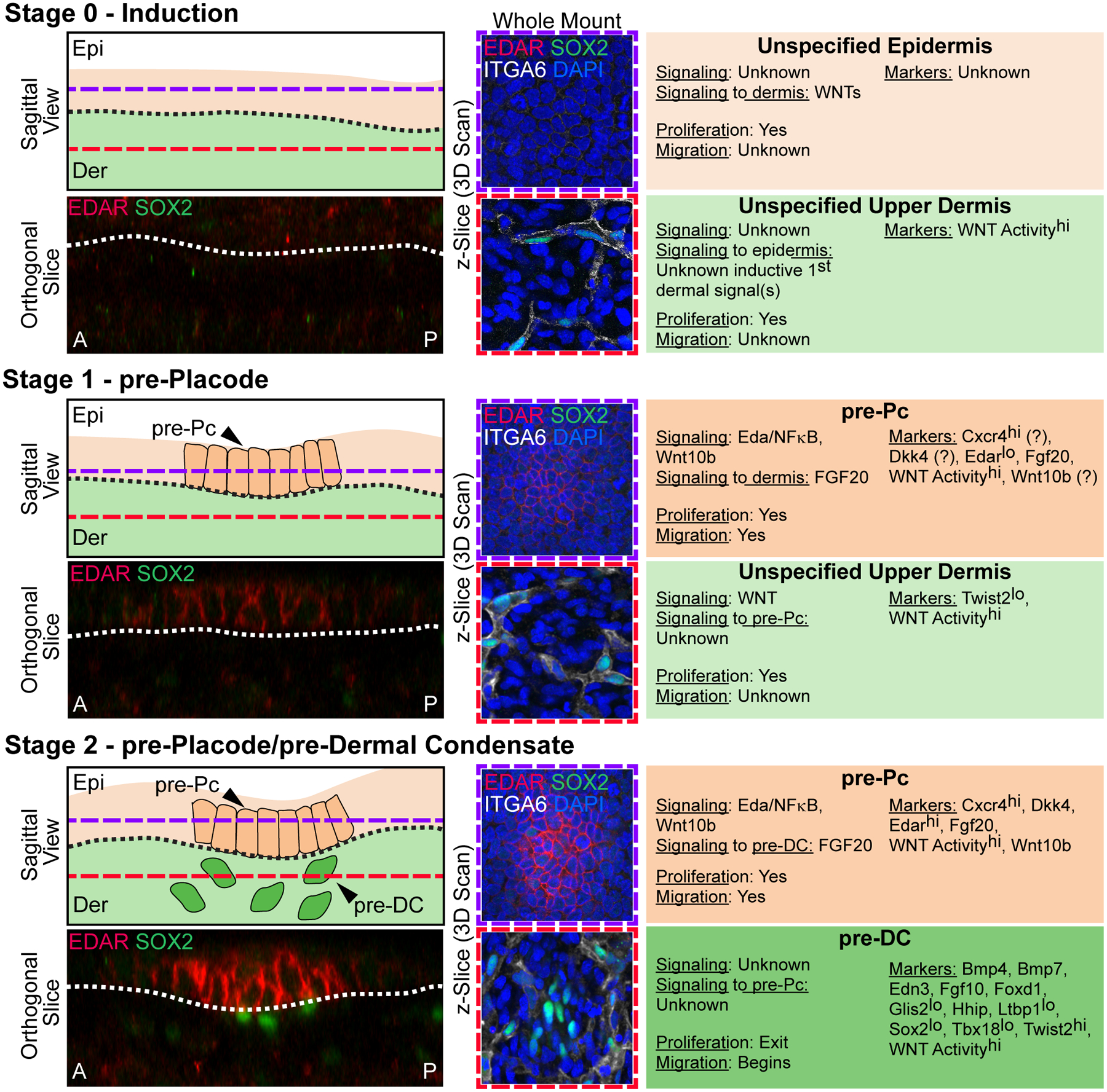
Figure 2. Stages 0–2 of Hair Follicle Morphogenesis.

Left top: Sagittal view schematic of HF morphogenesis stages. Purple dashed lines mark the epidermal Z-plane, and red dashed lines mark the dermal Z-plane of 3D-imaged confocal scans of whole mount immunofluorescence of E15.0 back skin. A and P denote anterior and posterior orientation of embryonic skin (head is left). Left bottom: Orthogonal slice from a 3D reconstruction of whole mount immunofluorescence for EDAR and SOX2. Middle: Whole mount immunofluorescence for EDAR, SOX2, and ITGA6 in the epithelial plane (top, purple frame), and dermal plane (bottom, red frame). DAPI marks all nuclei. Right: Description of autocrine and paracrine signaling, proliferation and migration status, and markers of the relevant epithelial and mesenchymal populations at each stage.
Stage 0 – Induction. The uniform unspecified multipotent epidermis resides over an unspecified dermis. EDAR is not expressed in the epidermal compartment and only SOX2+/ITGA6+ Schwann cells are present in the dermis. Widespread Wnt signaling activity in the upper dermis sets up HF induction by the critical, but still unknown “first dermal signal(s)”.
Stage 1 – pre-Placode. Emergence of placode precursors (pre-Pc), the fated “molecular placode”, in the epidermis over an unspecified dermis. The pre-Pc in the epidermal plane expresses EDAR. Only SOX2+/ ITGA6+ Schwann cells are present in the dermal compartment. Markers with question marks refer to known expression by in situ hybridization and/or protein staining at E13.5, in which the presence of recently discovered pre-DC (stage 2) cannot be ruled out.
Stage 2 – pre-Placode/pre-Dermal Condensate. pre-Pc in the epidermis over dermal condensate precursors (pre-DC) in the dermis. The pre-Pc in the epidermal plane expresses EDAR more strongly. Pre-DC emerge as low-level SOX2+/ITGA6− unclustered cells underneath pre-Pc.
