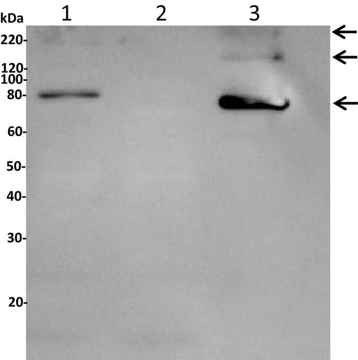
Fig. 3.

Gel blot analysis of AcTG‐1. For the immunological identification of AcTG‐1, an anti‐His antibody (1 : 3000) was used as a primary antibody and HRP‐conjugated goat anti‐mouse (1 : 5000) as a secondary antibody. The molecular weights of Magic Marker standards are indicated at the left margin. Lane 1: extract fraction; lane 2: elution fraction 1; and lane 3: elution fraction 3. The positions of positive AcTG‐1 bands are indicated by arrows.
