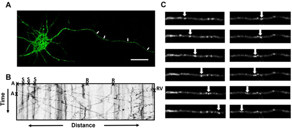
Figure 1.

Methods for testing compounds for their ability to affect axonal transport in vitro. (A) Representative image demonstrating successful transfection with pEGFP-n1-APP in rat primary cortical neurons. Arrows indicate membrane-bound organelles (MBOs), scale bar=20μm. (B) Kymograph generated from images captured at a rate of one frame every 2s for 3 min demonstrating movement of pEGFP-n1-APP labeled MBOs. MBOs are categorized in 1 of 4 ways: anterograde (A), retrograde (R), stationary (S), or Reversal (RV). (C) Representative frames demonstrating progression of MBOs moving in the anterograde and retrograde directions.
