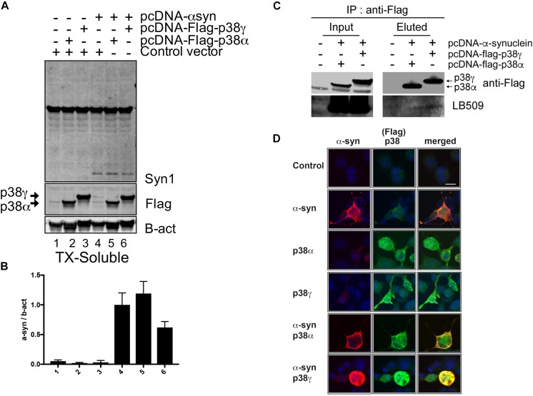
FIGURE 7.
Immunoprecipitation analysis between p38 and α-synuclein in a neuronal cell line. B103 neuroblastoma cells were transfected with pcDNA3, pcDNA-human-α-synuclein, pcDNA3-Flag-p38α, and pcDNA3-Flag-p38γ and analyzed by immunoblotting and immunocytochemistry. (A) After 48 h of transfection, cell lysates were analyzed by western blot and probed with an antibody against Flag to detect p38α, p38γ, and α-syn (Syn1); (B) levels of p38α, p38γ and α-syn were determined by densitometric quantification. (C) Co-immunoprecipitation of p38α, p38γ, and α-syn. p38α and p38γ were pulled down from transfected cell lysates and analyzed by western blot with an antibody against pathological α-syn (LB509) showing the interaction between α-syn and p38γ. (D) Representative images from double immunostaining for α-syn (red)/p38α (green) and α-syn (red)/p38γ (green). Strong colocalization (yellow) was observed in neuronal cells co-expressing α-syn and p38γ. Scale bar is 10 μm.

