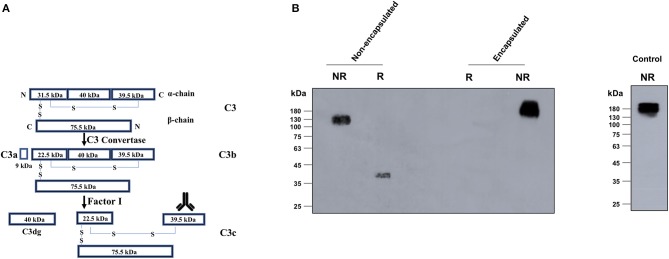
Figure 3.
Detection of the C3c processed fragment with encapsulated and non-encapsulated strains of Bacillus anthracis. (A) Schematic representation of the C3 processing during complement fixation depicting the cleavage of C3 into C3b, C3c, and C3d. C3b remains surface bound, whereas C3c is released in the fluid-phase media. (B) Non-encapsulated and encapsulated strains of B. anthracis were incubated in 10% normal human serum and C3c (138 kDa) detected in the supernatants by immunoblot assays using specific antibodies against the 40-kDa C-terminal fragment of alpha chain of C3c (marked by the antibody image in A). C3c formation was only observed with the non-encapsulated Sterne strain. The 140-kDa C3c (alpha and beta chains) protein and 40-kDa C3c (alpha chain C-terminus) were detected under non-reducing and reducing conditions, respectively, with the non-encapsulated B. anthracis Sterne strain. The unprocessed 189-kDa C3 protein from human serum was detected in the supernatants of the encapsulated virulent strain consistent with the same detected in the 1% normal human serum (positive control). R, reducing; NR, non-reducing.

