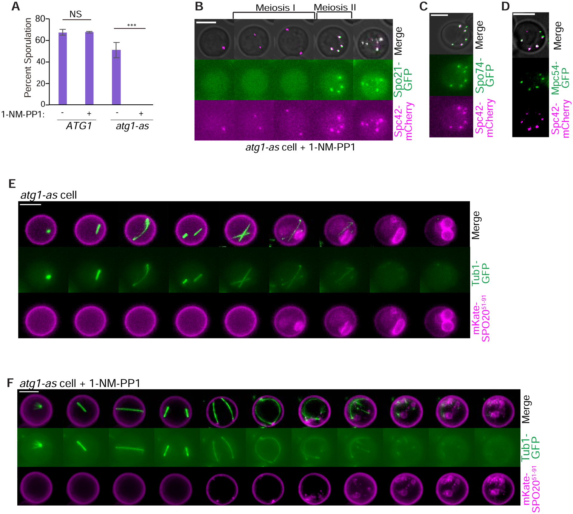
Figure 2: Autophagy inhibition disrupts gametogenesis.

A) Graph of percentage of wildtype and atg1-as cells (without Ndt80-IN) that sporulate with and without 1-NM-PP1 inhibitor added 10 hours after introduction into sporulation medium. NS = not significant. Asterisk indicates a statistically significant difference (n > 200 cells per strain; p<0.001; t test). B) Representative time-lapse images of an atg1-as cell undergoing synchronized meiosis in the presence of 1-NM-PP1 and β-estradiol addition. Scale Bar: 5μm. C, D) Similar to post-meiosis II images in part B but with additional fluorescently-labeled SPB components, as indicated. E-F) Representative time-lapse images of atg1-as cells undergoing synchronized meiosis in the absence (E) and presence (F) of 1-NM-PP1. Prospore membranes are marked with mKate-Spo2051–90. Scale Bars: 5μm.
