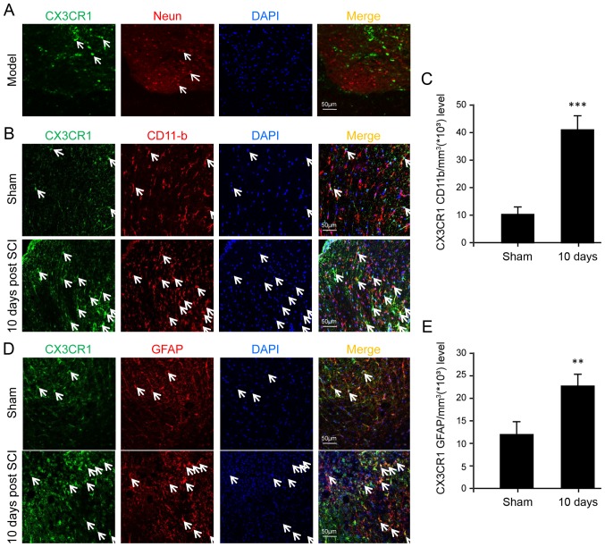
Figure 3.
Fluorescence immunostaining for cellular localization of CX3CR1 in the injured spinal cord after SCI. (A) Co-immunostaining of CX3CR1 (green) and the neuronal marker NeuN (red). Note that CX3CR1 was not localized in the NeuN positive cells (neurons). (B) Co-immunostaining of CX3CR1 (green) and the microglial marker CD11-B (red) after SCI. The arrows indicate the co-localization (yellow) of CX3CR1 and CD11-B. (C) The corresponding quantitative analysis is shown. (D) Co-immunostaining of CX3CR1 (green) and the astrocyte marker GFAP (red) in injured spinal cord tissues. The arrows indicate the co-localization (yellow) of CX3CR1 and GFAP. The corresponding quantitative analysis is shown in (E). The data was represented as the mean ± SD. **P<0.01 and ***P<0.001 vs. sham. Scale bar=50 μm. n=5. Me, methylprednisolone; ns, not significant; GFAP, glial fibrillary acidic protein; SCI, spinal cord injury; CX3CL1, C-X3-C motif chemokine ligand 1CX3CR1, C-X3-C motif chemokine receptor 1; DAPI, 4′,6-diamidino-2-phenylindole; CD, cluster of differentiation.

