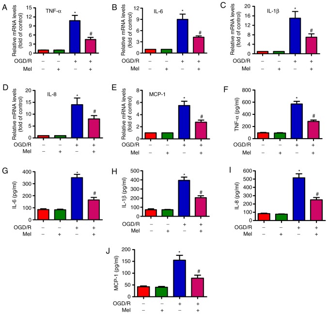
Figure 5.
Mel reduces the level of pro-inflammatory cytokines in OGD/R-insulted H9c2 cells. H9c2 cells were subjected to 4 h of OGD followed by reperfusion for 24 h. Mel (1 mM) was added to the culture medium at the initiation of reperfusion. (A–E) The mRNA expression levels of (A) TNF-α, (B) IL-6, (C) IL-1β, (D) IL-8 and (E) MCP-1 were analyzed by reverse transcription-quantitative PCR. (F–J) The release of (F) TNF-α, (G) IL-6, (H) IL-1β, (I) IL-8 and (J) MCP-1 in the supernatants was measured by ELISA. Data are expressed as the mean ± SD from three independent experiments. *P<0.05 vs. control; #P<0.05 vs. OGD/R. Mel, melatonin; OGD/R, oxygen-glucose deprivation/reperfusion; TNF-α, tumor necrosis factor-α; IL, interleukin; MCP, monocyte chemotactic protein.

