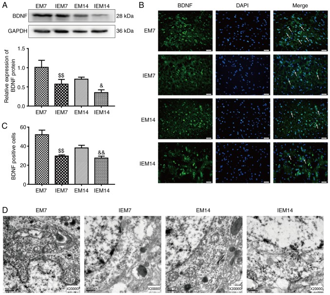
Figure 10.
Inhibition of the Wnt/β-catenin signaling pathway abolishes exercise-promoted BDNF expression and the morphology of cortical neurons in the ischemic penumbra after middle cerebral artery occlusion. (A) Protein expression levels and western blot analysis of BDNF. Data are presented as the mean ± SD (n=3). (B) Immunofluorescence of BDNF-positive cells in the ischemic penumbra. Scale bars=20 μm. (C) The number of BDNF-positive cells in the ischemic penumbra. Data are presented as the mean ± SD (n=6). (D) Transmission electron microscopy showed the neuron nucleus structures (n=3). Scale bars=0.5 μm. $$P<0.05 vs. the EM7 group; &P<0.05 and &&P<0.01 vs. the EM14 group. EM group, treadmill training model group. BDNF, brain derived natriuretic factor; SD, standard deviation.

