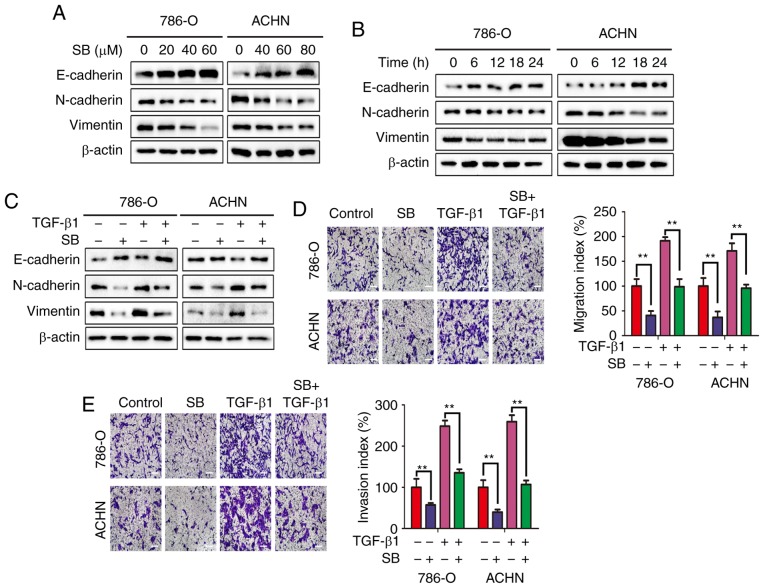
Figure 2.
SB suppresses EMT in RCC cells. Expression of E-cadherin, N-cadherin and vimentin in 786-O and ACHN was detected by western blotting following treatment with different concentrations of SB for (A) 24 h or (B) 60 μM SB for the indicated time intervals. (C) EMT markers were detected by western blot analysis following treatment with 60 μM SB and 5 ng/ml TGF-β1 for 24 h. β-actin was used as the loading control. (D) Transwell migration and (E) invasion assays were performed in cells treated with 60 μM SB and 5 ng/ml TGF-β1 for 24 h. DMSO treatment alone was used as the control. Magnification, ×100. Scale bar, 20 μm. **P<0.01. Results are shown as the mean ± standard deviation of three experimental repeats. RCC, renal cell carcinoma; EMT, epithelial-mesenchymal transition; SB, silibinin.

