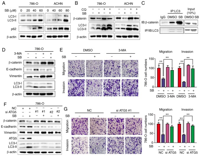
Figure 5.
SB induces inhibition of EMT via autophagy-dependent Wnt/β-catenin signaling in RCC cells. (A) Western blot analysis of LC3-I/II protein levels in 786-O and ACHN cells treated with different doses of SB. β-actin was used as the loading control. (B) 786-O and ACHN cells were treated with 60 μM of SB for 24 h in the presence of 50 μM CQ, and the protein expression levels of β-catenin and LC3-I/II were measured. β-actin was used as the loading control. (C) Co-immunoprecipitation of endogenous β-catenin and LC3 was assayed in 786-O cells following treatment with 60 μM SB. (D) 786-O cells were pretreated with 3 mM 3-MA for 1 h, followed by treatment with 60 μM of SB for 24 h. Western blot analysis was used to detect the expression levels of total β-catenin, E-cadherin, vimentin and LC3-I/II. (E) Transwell migration and invasion assays were performed on treated cells. Magnification, ×100. Scale bar, 20 μm. The experiment was repeated three times. **P<0.01. (F) 786-O cells were transfected with two siRNA sequences targeting ATG5 for 24 h and treated with DMSO or 60 μM SB for another 24 h. Western blot analysis was used to detect the expression levels of total β-catenin, E-cadherin, vimentin, ATG5 and LC3-I/II. β-actin was used as the loading control. (G) Transwell migration and invasion assays were performed on treated cells under similar conditions. Magnification, ×100. Scale bar, 20 μm. The experiment was repeated three times. **P<0.01. EMT, epithelial-mesenchymal transition; CQ, chloroquine; IB, immunoblotting; IP, immunoprecipitation; ATG5, autophagy-associated gene 5; SB, silibinin; RCC, renal cell carcinoma; 3-MA, 3-methyladenine; NC, negative control; E, epithelial.

