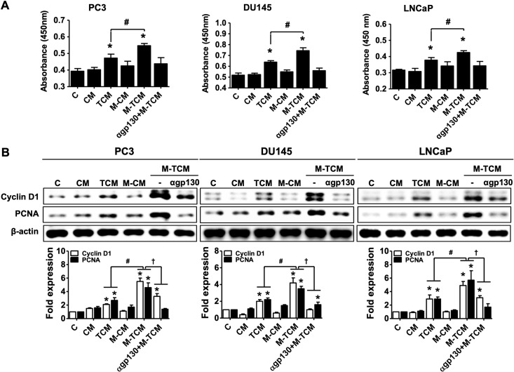
Fig 9. Proliferation of prostate cancer cells in response to conditioned medium of M2-like macrophages cultured with TCM.
To prepare the conditioned medium of M2-like macrophages, THP-1-derived macrophages were incubated in RPMI1640 media containing 20% TCM with or without pretreatment of gp130 antibody for 72 hr, and the culture supernatants were collected. The supernatants was named αgp130+M-TCM and M-TCM. Prostate cancer cells (PC3, DU145 and LNCaP) were cultured with CM, TCM, M-CM, M-TCM or αgp130+M-TCM for 24 hr. (A) Proliferation of the prostate cancer cells was measured by CCK-8 assay. (B) Cyclin D1 and PCNA were determined by western blot. Graph represent densitometric analysis (means of three independent western blot experiments). Data are means ± SD of three independent experiments. *p<0.05 versus untreated prostate cancer cells (C). #p<0.05 versus conditioned medium of RWPE-1 stimulated with T. vaginalis (TCM). †p<0.05 versus conditioned medium of RWPE-1 stimulated with T. vaginalis (TCM). CM: conditioned medium of RWPE-1 alone, TCM: conditioned medium of RWPE-1 stimulated with T. vaginalis, M-CM: conditioned medium of THP-1-derived macrophage stimulated with CM, M-TCM: conditioned medium of THP-1-derived macrophage stimulated with TCM, αgp130+M-TCM: conditioned medium of THP-1-derived macrophage stimulated with TCM after pretreatment of gp130 antibody.

