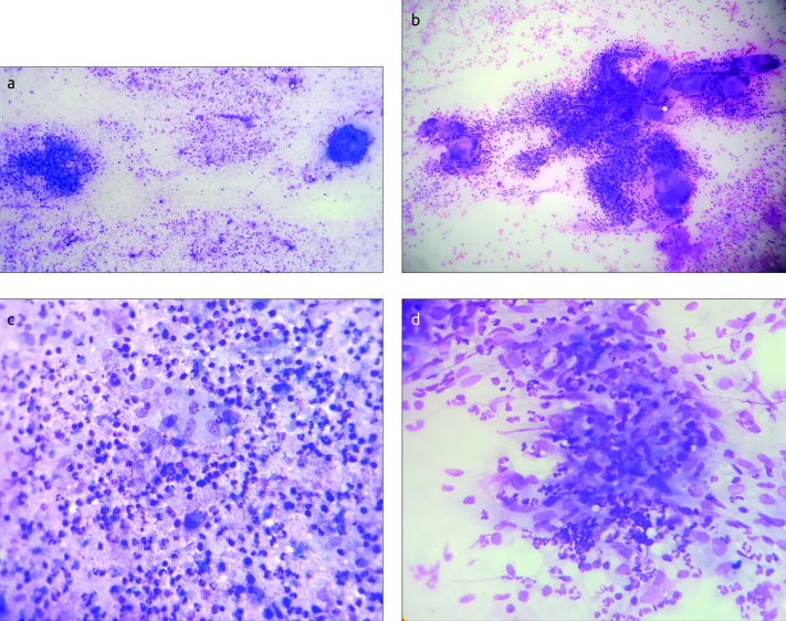
Figure 2. a–d.
(a) Smear show epithelioid granuloma, giant cell and suppurative background (Leishman stain ×100). (b): Smears show many epithelioid granulomas, giant cells and polymorphs (Leishman stain ×400). (c): Smears show many epithelioid granulomas, giant cells, polymorphs and few lymphocytes (Leishman stain ×400). (d): Smears show many epithelioid granulomas mixed with polymorphs (Leishman stain ×400)

