
Figure 5.
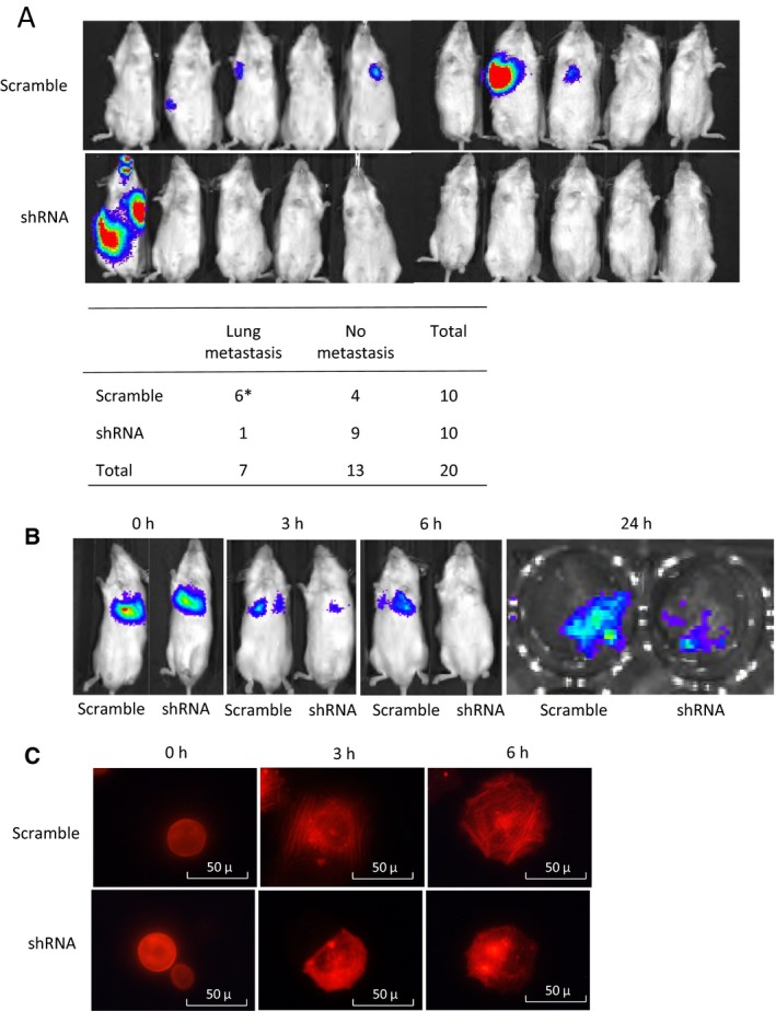
In vivo analysis of PAK1 silencing on OS metastasis. (A) Luminescence images of whole mice 4 weeks after tail‐vein injection of 143B PAK1‐shRNA#1 cells or scramble control cells. After an imaging analysis, mouse lungs were harvested and examined histologically to confirm the presence of pulmonary metastases. *One mouse in the scramble group developed micrometastasis in the lungs without detectable luminescence. Occurrence of metastasis was determined based on both luminescence and histopathological examination. The numbers of mice with or without metastases were compared between the shRNA and control groups (Fisher's exact test, P = 0.0286). (B) Luminescence images obtained early postinjection time points (0, 3, and 6 h) from mice injected with either the PAK1‐shRNA#1 or scramble control cells. The 24‐h image represents the luminescence image of the lung tissues harvested 24 h after tumor cell injection to illustrate tumor cell invasion of the lungs. (C) Representative fluorescence images of PAK1‐shRNA#1 and scramble cells at different time points (0, 3, and 6 h) after transduction with RFP‐actin in culture wells (20×). Experiments were replicated three times.