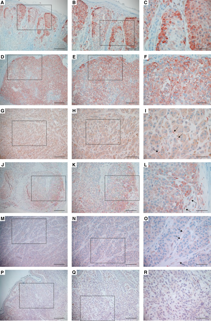
Figure 2.

Expression of P4HA1 protein in primary melanomas and nevi. (A–O) Examples of primary melanomas that show different levels of P4HA1 expression in melanoma cells. Staining was also detected in the fibroblasts surrounding the melanoma cells in some samples (I, L, O, marked with arrows). (P–R) P4HA1 expression in a benign nevus. Positive immunostaining is shown in red. (B, E, H, K, N, Q) show a higher magnification of the boxed area in (A, D, G, J, M, P, respectively) and (C, F, I, L, O, R) in (B, E, H, K, N, Q, respectively). Scale bars = 200 µm (A, D, G, J, M, P), 100 µm (B, E, H, K, N, Q), 50 µm (C, F, I, L, O, R).
