Figure 5.

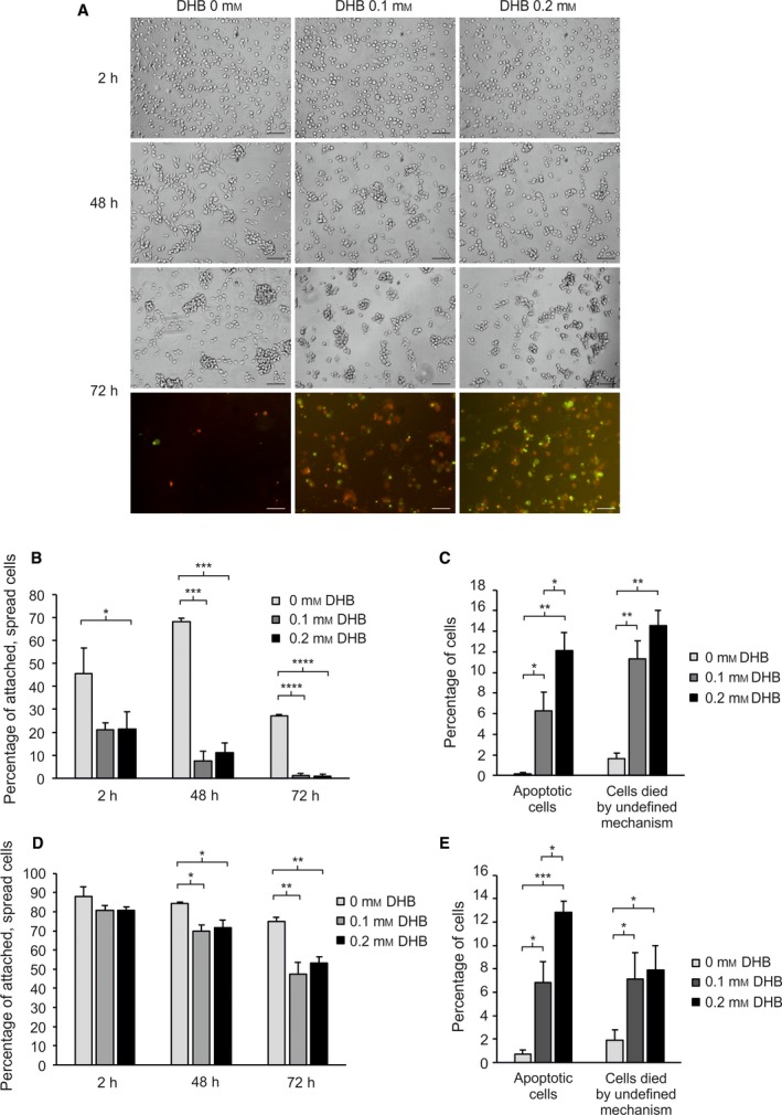
Effect of prolyl 4‐hydroxylase inhibition on cell adhesion and apoptosis/viability of SKMEL‐28 cells plated on uncoated or collagen‐I‐coated surfaces. (A) SKMEL‐28 cells were plated in serum‐free media without or with a prolyl 4‐hydroxylase inhibitor 3,4‐dihydroxybenzoic acid (DHB) and photographed after 2‐, 48‐, and 72‐h incubation. Both phase‐contrast and fluorescence images are shown after 72‐h incubation. Apoptotic cells (with activated caspase‐3/7) are seen in green and dead cells (stained with propidium iodide) in red. Apoptotic, dead cells are seen in yellow. (B–E) The percentage of attached and spread cells (B, D), apoptotic cells (green and yellow) (C, E), and cells died by undefined mechanism (red) (C, E) when incubated in serum‐free medium without or with DHB on uncoated (B, C) or collagen‐I‐coated (D, E) surfaces for 2, 24, and 72 h (B, D), or 72 h (C, E). Data are expressed as means ± SD of three replicates. *P < 0.05, **P < 0.01, ***P < 0.001, ****P < 0.0001. Scale bars = 100 µm.
