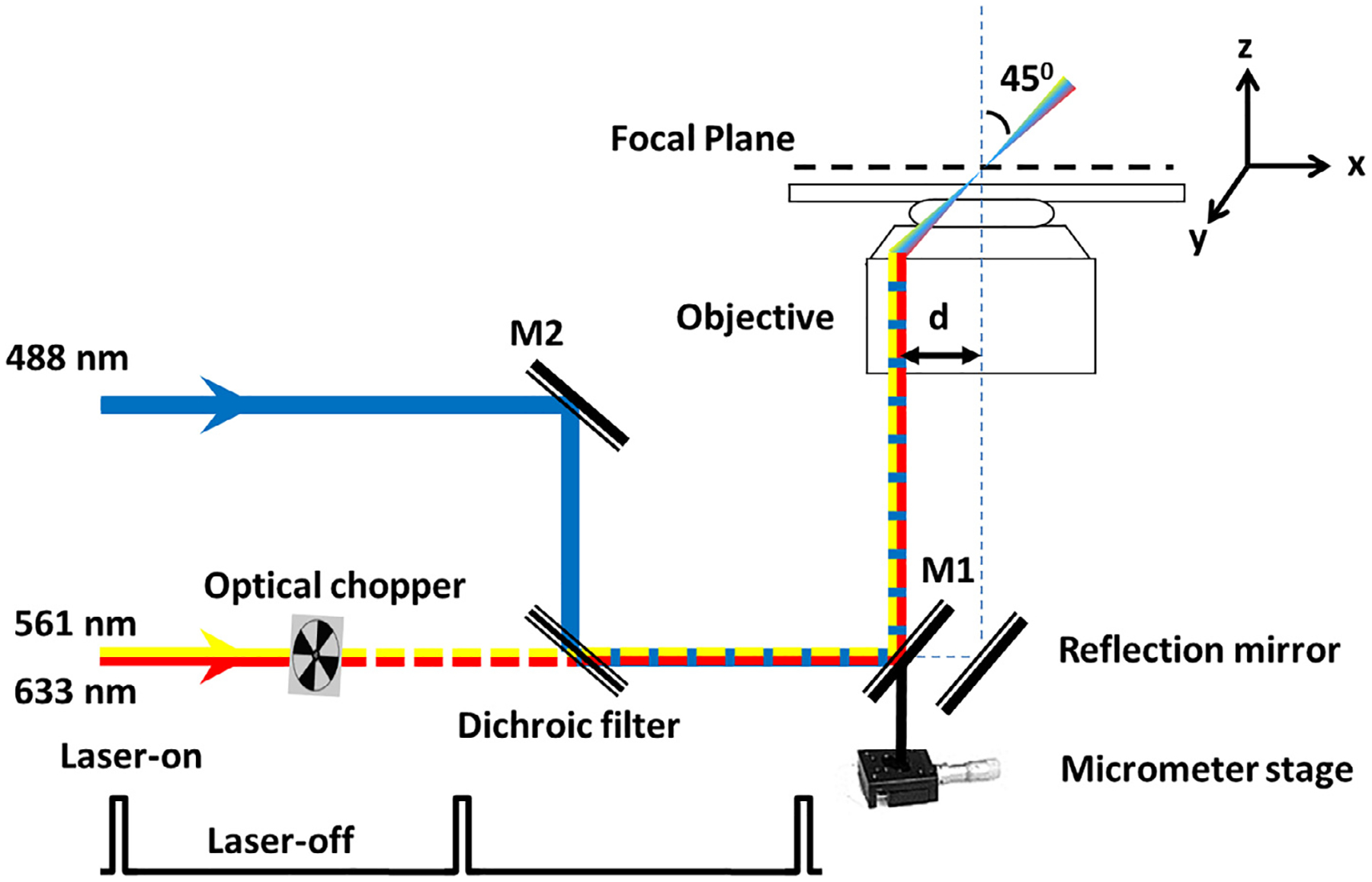
Fig. 6.

Optical schematic of the SPEED microscope setup [51]. A 488-nm, a 561-nm, and a 633-nm laser beams were co-aligned and then shifted together by ~237 μm (d) from the central optical axis of the objective to generate an inclined illumination volume at an angle of 45° to the perpendicular direction by using a micrometer stage. The 561-nm and 633-nm laser was chopped by an optical chopper to achieve an on-off laser mode with a laser-on time of 60 ms and a laser-off time of 140 ms. The longer laser-off time gives particles transiting the NPC sufficient time to escape from the illumination volume and for fresh fluorescent cargo to diffuse from the cytoplasm or the nucleus into the NPC.
