Figure 3.
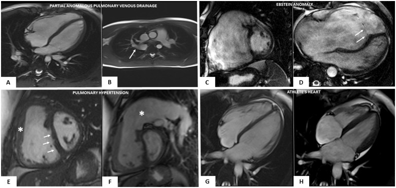
Cardiac magnetic resonance features of heart diseases mimicking right-dominant (classic) phenotypic variant of arrhythmogenic right ventricular cardiomyopathy. Partial anomalous pulmonary vein drainage (A and B): end-diastolic frame of cine cardiac magnetic resonance sequence in long-axis four-chamber view showing moderate right ventricular dilatation (A); cine sagittal view showing the anomalous drainage of the right pulmonary vein in the azygos vein (white arrow) (B). Ebstein anomaly (C and D): end-diastolic frame of cine cardiac magnetic resonance sequence in short-axis view showing a severe right ventricular enlargement due to a large ventricular ‘atrialization’ (C); end-diastolic frame of cine cardiac magnetic resonance sequence in four-chamber view showing a significant apical displacement of the septal leaflet of the tricuspid valve (white arrows) (D). Arterial pulmonary hypertension (E and F): end-diastolic frames of cine cardiac magnetic resonance sequence in short-axis view showing increase of the right ventricular wall thickness (white asterisk) (E), flattening of the interventricular septum (white arrows) (E), and massive pulmonary artery dilatation (white asterisk) (F). Athlete’s heart (G and H): end-diastolic (G) and systolic (H) frames of cine cardiac magnetic resonance sequence in four-chamber view evidencing biventricular dilatation (end-diastolic volume 122 mL/m2) and normal systolic function (ejection fraction 64%), in the absence of wall motion abnormalities (not shown).

