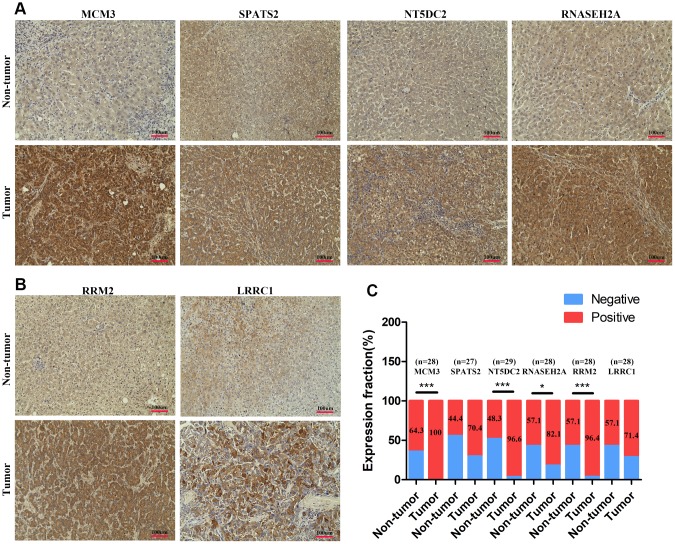
Figure 5.
The expression levels of MCM3, SPATS2, NT5DC2, RNASEH2A, LRRC1, and RRM2 in HCC tissues. (A–B) Immunohistochemical staining analysed expression levels of MCM3, SPATS2, NT5DC2, RNASEH2A, LRRC1, and RRM2 in HCC and non-tumor tissues. (C) Positive expression percentage of the six genes in HCC and non-tumor tissues was showed. Fewer than 30 samples due to de-fragmentation. *P < 0.05; **P < 0.01; ***P < 0.001.

