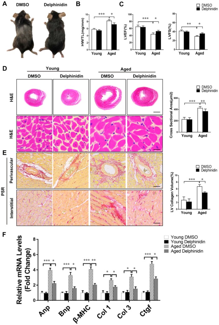
Figure 8.
Delphinidin reduced cardiac hypertrophy in aged mice. (A) Representative gross morphology of young and aged mice administered delphinidin and DMSO. (B) Statistical analysis of differences in the heart weight/tibia length (HW/TL) ratio (n=6). (C) Left ventricular ejection fraction and fractional shortening of young and aged mice administered delphinidin and DMSO (n=6). (D) Left, H&E staining was performed to assess hypertrophic growth of the hearts of young and aged mice administered with delphinidin and DMSO. Right, Statistical analysis of differences in cardiomyocyte size (n=6). (E) Left, Representative PSR staining of histological sections of the LV (n=6). Right, Statistical analysis of differences in cardiac fibrosis. (F) Quantitative real-time PCR (qRT-PCR) was performed to analyze the mRNA levels of hypertrophic genes and fibrosis genes (n=5). In B–F, *p<0.05, **p<0.01, ***p<0.001.

