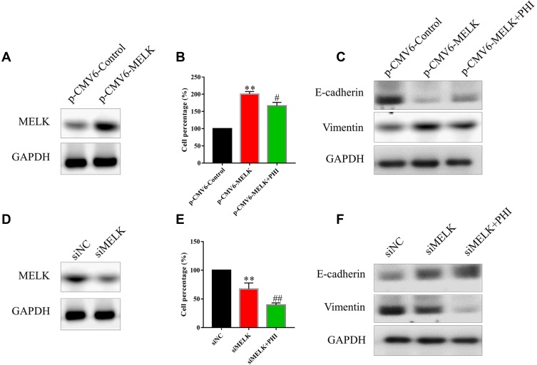
Figure 6.
PHI inhibited cell proliferation and EMT by inhibiting MELK. (A) Overexpression of MELK by p-CMV6-Myc-MELK in PANC-1 cells. MELK protein levels were assessed 24 h after transfer of p-CMV6-MELK. (B) Overexpression of MELK increased cell proliferation in PANC-1 cells. (C) Overexpression of MELK increased EMT in PANC-1 cells. (D) Knockdown of MELK expression by siMELK in PANC-1 cells. MELK protein levels were assessed 48 h after transfer of siMELK. (E) The silencing of MELK decreased cell proliferation in PANC-1 cells. (F) The silencing of MELK decreased EMT in PANC-1 cells. **p<0.01, vs p-CMV6-Control or siNC group, #p<0.05 or ##p<0.01, vs p-CMV6-MELK or siMELK group.

