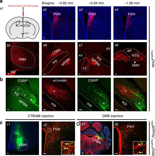
Fig. 5. Projections of PVHTrkB neurons.
a AAV2-CAG-FLEX-tdTomato was unilaterally injected into the PVH of Ntrk2CreER/+ mice (a1). After the mice were treated with tamoxifen, tdTomato-labeled PVHTrkB neurons were present throughout the rostral-caudal axis (a2–a4). Axonal terminals of these neurons were found in the VMH, LPBN, MPBN, LC, NTS and DMV (a5–a8). b The LPBN includes the external (e), central (c), and ventral (v) compartments. Axonal terminals of PVHTrkB neurons are mainly in the central compartment, which is next to the CGRP-marked external compartment. c, d Retrograde tracers, CTB488 (250 nl) or GRB (200 nl), were unilaterally injected into the VMH (c) or LPBN (d) of tamoxifen-treated Ntrk2CreER/+;Rosa26Ai9/+ mice and labeled some tdTomato-expressing PVHTrkB neurons. AP area postrema; DMV dorsal motor nucleus of the vagus; ME median eminence; LC locus coeruleus; LPBN lateral parabrachial nucleus; MPBN medial parabrachial nucleus; NTS nucleus tractus solitaris; PVH paraventricular hypothalamus; scp superior cerebellar peduncle; VMH ventromedial hypothalamus. Scale bars represent 200 μm in d1 and 50 μm in other panels. Source data are provided as a Source Data file.

