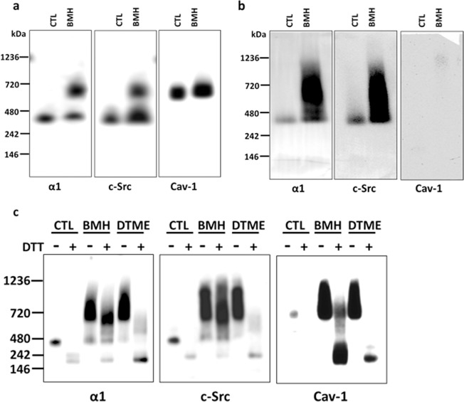
Figure 3.
BN-PAGE analysis of LLC-PK1 and C2-9 cells: The α1 subunit and c-Src form protein-protein complex under native condition that is not dependent on cav-1. Crosslinking and preparation of whole cell lysate with Native-PAGE sample buffer were performed as described for BN-PAGE in the Materials and Methods. NativeMark unstained protein standard was located by Ponceau S staining after transferring to PVDF membrane. Control (CTL, with mocking crosslinking process without crosslinkers) and BMH- or DTME-crosslinked samples (25 μg protein/sample) were processed side-by-side and immunoblotted for the α1 subunit, c-Src, and cav-1, respectively. For (a,b), the same CTL and crosslinked samples were separated into 3 groups (each group contains one CTL and crosslinked samples and separated by NativeMark protein standard between groups) and run in the same gels. After transferring, the PVDF membrane was cut into the 3 groups and immunoblotted against each antibody individually. (a) BN-PAGE analysis of control (CTL) and BMH-crosslinked samples of LLC-PK1 cells. (b) BN-PAGE analysis of control (CTL) and BMH-crosslinked samples cav-1 depleted C2-9 cells. (c) Each sample (CTL, BMH- and DTME-crosslinked sample) of LLC-PK1 cells was treated with or without DTT/SDS (100 mM DTT with 1% SDS, final concentration, respectively). For DTT/SDS treatment, samples were heated at 60 °C for 30 min (for the α1 subunit) or 95 °C for 5 min (for c-Src and cav-1). FluorChem M imager system (ProteinSimple) was used to detect blot signals. n = 3–4.

