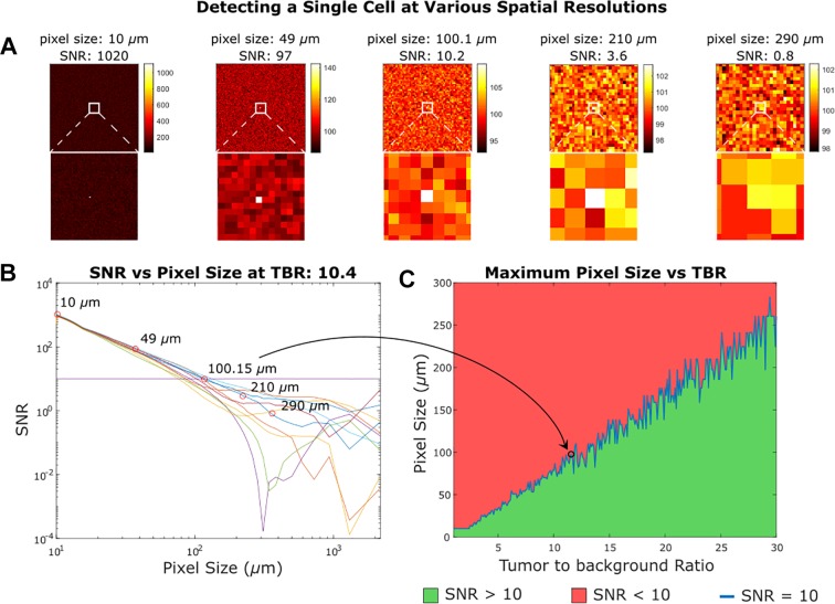
Figure 7.
Detecting a single cell at various spatial resolutions. In (A) we see the results of imaging a 10-micron diameter cell in healthy tissue with some noise and a tumor to background ratio of 10.4. We can still determine the location of the cell at the center of the imager as long as the signal to noise ratio (SNR) is greater than 10. Detecting a single cell does not require subcellular resolution contingent on the notion that the single cells are sparsely distributed and have a large tumor to background ratio. In (B) we see the SNR over a range of pixel sizes for 10 randomly generated samples given a tumor to background ratio (TBR) of 10.4. In (C) we plot the largest pixel that can detect a single cell at a given TBR with SNR greater than 10. The plot is not smooth because it is based on randomly generated samples with arbitrary noise.

