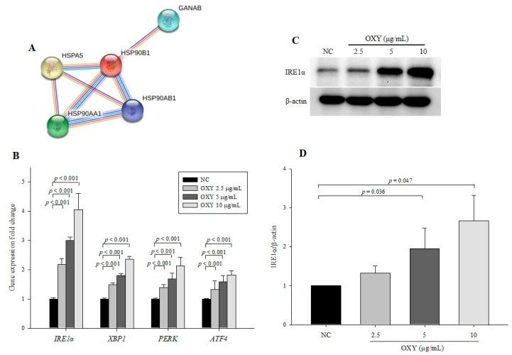
Figure 7.
Effect of OXY on the expression levels of endoplasmic reticulum (ER) stress signaling pathway-related genes and proteins. Protein–protein interaction images were displayed using STRING (A). Expression levels of IRE1α, XBP1, PERK, and ATF4 were determined by qPCR (B). The expression level of the IRE1α protein was measured by Western blotting (C) and quantified (D). From each lysate, equal amounts of protein were loaded on a separate gel, blotted for actin, and this signal was used for determining the ratio of the protein of interest/actin as displayed in the figure. The cropped blots are representative of three independent experiments. The full-length blots are shown in Supplementary Figure S5. Each value indicates the mean ± SD of three independent experiments performed.

