Figure 2.
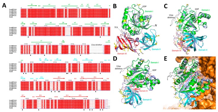
Protein structural modelling of LreEF1A4. (A) Amino acid sequence alignment of LreEF1A4 with other homologs, including Lilium regale LreEF1A1–3, Arabidopsis AteEF1A1, and mammal 5LZS. The secondary structure is displayed above the sequences in green (domain I), cyan (domain II), and red (domain III). Amino acid substitution sites within LreEF1A1–4 are indicated with solid triangle. (B–D) Structural superimposition of LreEF1A4 model with mammalian eEFA1 5LZS (blue) in different angles. (E) Spatial orientation of LreEF1A4 in complex with ribosomal proteins (orange). Domain I, II, and III are highlighted in green, cyan, and red, respectively. Amino acid substitution sites within LreEF1A1–4 are shown in yellow sticks.

