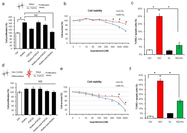
Figure 1.
KL (α-klotho) attenuated isoproterenol-induced cell death in H9C2 cardiomyocytes. (a) Un-differentiated H9c2 cells were cultured with KL (200 ng/mL), fibroblast growth factor-23 (FGF23) (100 ng/mL), KL (200 ng/mL) plus FGF23 (100 ng/mL), KL (200 ng/mL) + anti-FGF23 (1 μg/mL), or KL (200 ng/mL) plus anti-KL (1 μg/mL) for 3 days. Cell proliferation activity was measured on day 3 using the Cell counting kit-8 (CCK-8) kit. (b) Un-differentiated H9c2 cells were cultured with different doses of isoproterenol in the presence or absence of KL (200 ng/mL). Cell viability was measured 48 h after the treatments using the CCK-8 kit. (c) TUNEL-positive cells were analyzed after immunofluorescence staining of cells treated with isoproterenol (1000 nM), KL (200 ng/mL), or isoproterenol (1000 nM) plus KL (200 ng/mL). Cells were fixed and stained 24 h post-treatment. (d) H9c2 cells were cultured in retinoic acid (RA) and 1% FBS for 5 days to induce differentiation and maturation. Differentiated H9c2 cells were then cultured with KL (200 ng/mL), FGF23 (100 ng/mL), KL (200 ng/mL) plus FGF23 (100 ng/mL), KL (200 ng/mL) + anti-FGF23 (1 μg/mL), or KL (200 ng/mL) plus anti-KL (1 μg/mL) for 3 days. Cell proliferation activity was measured on day 3 using the CCK-8 kit. (e) Differentiated H9c2 cells were cultured with different doses of isoproterenol in the presence or absence of KL (200 ng/mL). Cell viability was measured 48 h after the treatments using the CCK-8 kit. (f) TUNEL-positive cells were analyzed after immunofluorescence staining of cells treated with isoproterenol (1000 nM), KL (200 ng/mL), or isoproterenol (1000 nM) plus KL (200 ng/mL). Cells were fixed and stained 24 h post-treatment. In all graphs, * indicates p < 0.05. Data are representative of three independent experiments.

